3/30/2020 Ver. A
ELISA Disease Detection
Modeling Antigen and Antibody Detection

Note to Teachers
STOP: Read this slide before proceeding, then delete it.
This activity is designed for students to work though on their own. Please adapt it to fit the needs of
your course.
Make it relevant — pick a disease that is of particular interest to your students, and have students
model the use of ELISA for its detection. A coronavirus scenario is presented in the antibody
detection ELISA slides.
Make it accessible — instead of printing and cutting out shapes, students can click and drag the
shapes to create a digital model; they may also choose to draw their own model.
Add an assessment — students can record and narrate their models using Flipgrid or Stop
Motion Studio apps. Assessment question slides have a green border along the top edge.
Copyright 2020 Bio-Rad Laboratories, Inc. All content and images contained in this presentation may be used for educational purposes only and may not be
distributed. Bio-Rad is a trademark of Bio-Rad Laboratories, Inc. in certain jurisdictions. All trademarks used herein are the property of their respective owner.
explorer.bio-rad.com
2

Enzyme-Linked ImmunoSorbant Assay (ELISA)
ELISAs use antibodies to detect proteins in a patient’s
sample.
Many diseases and conditions can be detected and
diagnosed with ELISAs, including:
• Influenza
•HIV
• SARS
• Zika virus
• West Nile virus
• Lyme Disease
Researchers are also developing ELISAs that can be
used to diagnose current or past coronavirus (SARS-
CoV-2) infection.
explorer.bio-rad.com
3
Novel Coronavirus SARS-CoV-2
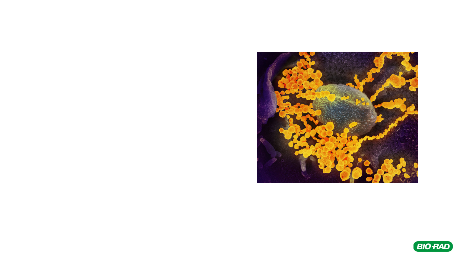
This scanning electron microscope image shows SARS-CoV-2
(round gold objects) emerging from the surface of cells cultured
in the lab. SARS-CoV-2, also known as 2019-nCoV, is the virus
that causes COVID-19. The virus shown was isolated from a
patient in the U.S. Credit: NIAID-RML

Antigen-Antibody Interactions
Pollen, bacteria, viruses, and other foreign molecules are
seen by your body as invaders, and they create an
immune response. These foreign invaders are called
antigens.
Your immune system makes antibodies in response to
antigens. The antibodies bind antigens, flagging them for
destruction by immune cells.
Antibodies have two regions: variable and constant. Each
tip of the “Y” in the variable region is highly specific and
binds to only one particular antigen. The constant region is
the same for every antibody of the same type (there are 5
different types of antibodies).
Antibodies can also be produced by injecting an animal
with antigen - disease agents or even antibodies from a
different type of animal.
explorer.bio-rad.com
4
Antibody
Constant
Region
Variable
Region
Antigen

Infection Timeline — Antigens and Antibodies
5
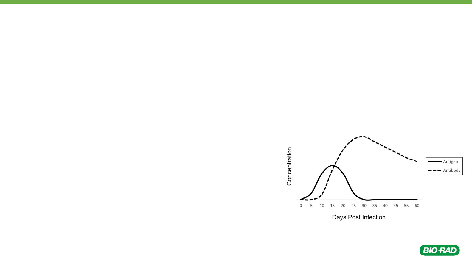
After infecting a person, a virus
multiplies in the body and the
concentration of viral antigens
increases.
Soon after infection, the immune
system kicks in and starts producing
antibodies.
As the antibody concentration
increases, antibodies bind to viruses
and target them for destruction by
the immune system.
Eventually, the viral antigen
concentration drops, the infection is
cleared, and the person recovers.

Antigen Detection ELISA Components
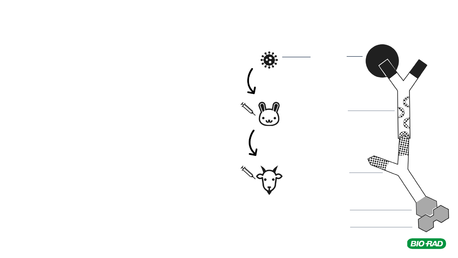
● Antigen — in an antigen detection ELISA, the
patient sample is tested for the presence of
antigens from viruses, bacteria, etc.
● Primary antibody — binds to the antigen
○ can be produced in a lab by injecting the target
antigen into an animal and then harvesting the
serum
● Secondary antibody — binds to the constant
region of the primary antibody
○ made by injecting the primary antibodies from
one animal into a different animal
○ secondary antibodies are attached to an
enzyme which catalyzes a color change when
substrate is added
● Substrate — changes color in the presence of the
enzyme, indicating a positive result
6
Primary
Antibody
(rabbit anti-virus)
Secondary
Antibody
(goat anti-rabbit)
Enzyme
Antigen
(virus)
explorer.bio-rad.com
Substrate

Influenza Antigen Detection
7
explorer.bio-rad.com
Hemagglutinin
(HA)
Influenza binds to target cells via the
hemagglutinin glycoprotein, HA. Scientists can
create antibodies that detect and bind to
influenza by injecting HA into an animal.
cross-section of influenza virus
https://www.cdc.gov/flu/resource-
center/freeresources/graphics/images.htm
Primary
Antibody
(rabbit anti-influenza
HA)
Secondary
Antibody
(goat anti-rabbit)
Enzyme
Antigen
(influenza HA)
Substrate

Model an Antigen Detection ELISA
1. Print out the paper model shapes on the LAST slide in this
presentation (click on the last slide, then go to File > Print, and
change Print All Slides to Print Current Slide).
2. Cut out each shape.
3. Grab one more blank sheet of paper.
4. Gather the following shapes for the first activity:
• 2 x primary antibodies with solid black variable region
• 5 x secondary antibodies
• 1 of each antigen shape (triangle, circle, rectangle)
• 6 x substrate
-OR-
Go to the second to last slide (Digital ELISA Model) and build
your model digitally: click and drag to move the shapes into the
well.
explorer.bio-rad.com
9
Primary
Antibody
Secondary
Antibody
SubstrateAntigens

Note: the pattern on the ends of the
primary antibodies match specific
patterns on the antigens.
The pattern and shape of the
secondary antibodies matches the
pattern and shape on the constant
region of the primary antibody.
These patterns and shapes
represent both the specificity and
location of antigen-antibody, and
antibody-antibody binding.
explorer.bio-rad.com
10
Model an Antigen Detection ELISA

Antigen Detection ELISA Model – Setup
An ELISA is typically performed in a plastic microstrip
plate that has 12 wells.
Each strip has wells for a positive control, a negative
control, and two patient samples, each done in triplicate
(4 samples x 3 wells each = 12 wells).
The blank sheet of paper represents a well in a
microstrip plate. You will model an ELISA on your
paper well.
As you read through the steps in an ELISA, use your
paper shapes to model the steps. The actual ELISA
steps are explained at the top, and the model steps are
explained underneath.
explorer.bio-rad.com
11
The blank paper
represents one well in
the microstrip plate

Antigen Detection ELISA – Step 1
Assay Step 1 — Bind antigen to the well
a. Add the patient sample to the well. This sample contains many
different proteins that may or may not contain the antigens that you
are trying to detect. These proteins bind non-specifically (adsorb) to
the plastic well, due to hydrophobic interactions.
b. Incubate the sample for 5 minutes, then tap out the fluid.
c. Add wash buffer to the well to rinse out anything that isn’t bound, and
to block the inner surface of the well. This prevents anything from
binding non-specifically in future steps.
d. Repeat the wash step.
Model Step 1
a. Add the antigens around the bottom edge of your blank paper “well”.
These represent the proteins / antigens that might be present in any
given patient sample.
explorer.bio-rad.com
12

Antigen Detection ELISA – Step 2
Assay Step 2 — Bind primary antibody to antigen
a. Add the primary antibodies to the well. The antibodies bind only to a
specific antigen out of the many that may be bound to the well. For
example, an anti-HIV antibody would only bind to HIV antigen. This
antigen-antibody interaction is specific and strong.
b. Incubate the sample for 5 minutes, then tap out the fluid.
c. Add wash buffer to the well to rinse out anything that isn’t bound. If
the primary antibody did not bind to any antigen, then it will be
washed away in this step.
d. Repeat the wash step.
Model Step 2
a. Add the primary antibodies into your well. Overlay the antibody so
that the black regions align. This represents specific antibody-antigen
binding.
b. Model the wash step by removing the unbound primary antibody.
explorer.bio-rad.com
13

Antigen Detection ELISA – Step 3
Assay Step 3 — Bind secondary antibody
a. Add the enzyme-linked secondary antibodies to the well. These
antibodies bind tightly to any primary antibodies that are present. The
secondary antibodies are covalently linked to an enzyme, horseradish
peroxidase, which will catalyze a reaction with a substrate to produce
a color change.
b. Incubate the sample for 5 minutes, then tap out the fluid.
c. Add wash buffer to the well to rinse out any secondary antibodies that
did not bind.
d. Repeat the wash step 2 more times (3 times total).
Model Step 3
a. Add the secondary antibodies into your well. Overlay the secondary
antibody so that the patterns align with the primary antibody.
b. Model the wash step by removing the unbound secondary antibody.
explorer.bio-rad.com
14

Antigen Detection ELISA – Step 4
Assay Step 4 — Add enzyme substrate for detection
a. Add the substrate to the well, wait 5 minutes, and evaluate results. If
the antigen was present, then the primary and secondary antibodies
bound and the well will turn blue. If there was no antigen, then no
primary or secondary antibodies bound, so the well will remain clear.
Model Step 4
a. Add the substrate into your well. Align the substrate with the enzyme
on the secondary antibody.
b. At this point, the enzyme catalyzes a reaction where the substrate
turns blue - a positive result.
explorer.bio-rad.com
15

Questions — Antigen Detection ELISA
1. In a typical ELISA, lab technicians use separate wells for positive controls, negative controls, and patient
samples. You modeled the well of a positive patient sample.
a) Use your shapes to model a positive control. Describe the necessary components and steps.
b) Use your shapes to model a negative patient sample. Describe the necessary components and steps.
explorer.bio-rad.com
16
2. Examine this graph of antigen and antibody concentrations
over time.
a) Could you use an antigen detection ELISA to accurately
diagnose a patient on day 35? Explain your answer.
b) Antigen detection ELISAs detect viral proteins. Viral DNA
or RNA can be detected using a different technique called
PCR (polymerase chain reaction). Using the graph,
explain when a viral PCR test would be most useful.

Questions — ELISA Design
3. Refer to the diagram, and notice where the primary
antibody binds antigen and secondary antibodies.
a) Could goat anti-rabbit secondary antibodies be
used to detect two different types of rabbit
antibodies? Explain your answer.
b) If HIV antigen was injected into the rabbit instead
of influenza, could the same secondary
antibodies (goat anti-rabbit) be used to detect
both rabbit anti-influenza antibodies AND rabbit
anti-HIV antibodies? Explain your answer.
explorer.bio-rad.com
17
Primary
Antibody
(rabbit anti-influenza
HA)
Secondary
Antibody
(goat anti-rabbit)
Enzyme
Antigen
(Influenza HA)
Substrate

Question — ELISA Design
4. You modeled an indirect ELISA, which uses enzyme-linked
secondary antibodies. In a direct ELISA, the enzyme is linked to
the primary antibody instead, so no secondary antibody is required.
Look at your model and think about what might happen if the
enzyme was attached to the primary antibody directly, so that a
secondary antibody was not needed.
a) Would you still get a blue color change when the substrate was
added? Explain your answer.
b) How might the signal intensity (amount of color change)
compare to the indirect ELISA that you modeled first?
c) What might be one advantage to using an enzyme-linked
secondary antibody?
explorer.bio-rad.com
18

Antibody Detection ELISA Overview
ELISAs can also use purified antigen to detect
antibodies in a patient’s serum. The presence of viral
antibodies in a patient sample indicates a current or
previous infection.
Coronavirus (SARS-CoV-2) binds to target host cells
via its spike glycoprotein, S. When someone is
infected with coronavirus, their immune system makes
antibodies to viral proteins, including S protein.
Scientists are developing ELISAs that use purified S
protein as the antigen to detect the presence of
coronavirus antibodies in a patient’s serum. ELISAs
that detect other viral proteins are being researched as
well.
explorer.bio-rad.com
19
Spike
Glycoprotein
(S)

Coronavirus Antibody Detection ELISA
20
Coronavirus
infects person
explorer.bio-rad.com
Coronavirus Infection
Coronavirus Antibody Detection ELISA
Person makes
antibodies against
coronavirus
Antigen
(lab-purified coronavirus S
protein)
Patient Sample
(anti-coronavirus antibodies
will be present in sample if
patient was infected)
Secondary Antibody,
goat anti-human
(binds if anti-coronavirus human
antibodies are present in the sample)
Enzyme
Substrate

Questions — Antibody Detection ELISA
explorer.bio-rad.com
22
5. In an antibody detection ELISA, lab-purified antigen (coronavirus S protein, for example) is added to the
wells first. Model this step using the solid black circles to represent the coronavirus S protein antigens.
How is this step different than the first step in an antigen detection ELISA?
6. After antigen is added, the wells are washed. Why is the wash step important? Give two reasons.
7. Next, the patient sample is added. Patient samples contain a variety of antibodies. If the patient has
been infected with coronavirus, the sample will also contain antibodies to the coronavirus S protein.
Model this step for a patient who has antibodies to coronavirus. Describe the necessary components
and steps.
8. An enzyme-linked secondary antibody is added next. Model this step and describe the necessary
components and steps.
9. Finally, the substrate is added. Model this step. If the sample turns blue (a positive result) the patient
sample contains antibodies to coronavirus. Does this necessarily indicate an ongoing infection? Explain
your answer.

Questions — Antibody Detection ELISA
explorer.bio-rad.com
23
10. Use your shapes to model the following for an antibody
detection ELISA:
a) The positive control
b) The negative control
c) A negative patient sample
11. According to this graph, could you use an antibody
detection ELISA to accurately diagnose a patient 35
days after exposure? Explain your answer.

explorer.bio-rad.com
24
ELISA well
Digital ELISA Model

explorer.bio-rad.com
25


